Retinal Evaluation & Treatment at Vinayaka Nethralaya
Preserving your central vision through thorough retinal assessment & timely treatment
The retina is the thin layer of light-sensitive tissue at the back of the eye that makes vision possible. When it is affected by disease — whether from diabetes, age-related changes, vascular problems, or structural issues — the consequences for vision can be serious and in some cases permanent. Many retinal conditions develop gradually and without noticeable symptoms in their early stages, which means that by the time a patient becomes aware of a visual change, meaningful damage may already have occurred. Others — such as retinal detachment or the sudden onset of wet macular degeneration — can cause rapid and severe vision loss if not assessed and treated without delay. At Vinayaka Nethralaya, retinal evaluation involves a thorough dilated fundus examination combined with appropriate diagnostic imaging to build an accurate picture of the retina's health. Treatment is then guided by the specific findings, with the aim of halting further deterioration and preserving the best possible vision for each patient.
Dilated fundus examination and retinal imaging assessment
Clear, affordable consultation fees
Experienced ophthalmologist with over four decades of clinical practice
Retinal evaluation & diagnostic imaging
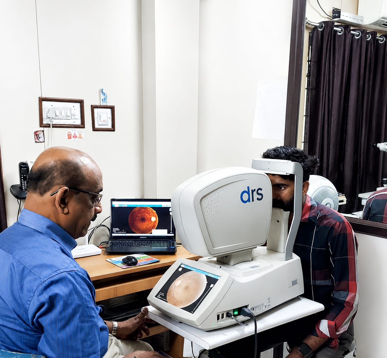
Each retinal assessment begins with a dilated fundus examination to allow a direct and detailed view of the retina, macula, optic nerve, and peripheral retinal structures. Where vascular assessment is required, fluorescein angiography is used to evaluate retinal circulation and identify areas of leakage or poor blood supply. Widefield fundus photography extends the view to the peripheral retina, where breaks, degeneration, and vascular changes can occur without any central visual symptoms.
Diabetic retinopathy & retinal vascular disease
Diabetic retinopathy develops when prolonged high blood sugar causes damage to the small blood vessels supplying the retina — leading progressively to fluid leakage, areas of poor circulation, and in advanced cases the growth of abnormal new vessels that can bleed or cause tractional damage to the retina. All patients with diabetes should have their retina examined regularly even in the absence of visual symptoms, as significant retinopathy can be present before any change in vision is noticed. Treatment for diabetic macular oedema and proliferative retinopathy includes intravitreal injections and targeted laser therapy, with the approach tailored to the stage and pattern of disease. Retinal vein occlusion causing macular swelling is managed along similar lines.
Age-related macular degeneration & macular conditions
Age-related macular degeneration is among the most frequent causes of central vision loss in adults over fifty. The dry form progresses gradually as the cells of the central retina deteriorate over time. The wet form — caused by abnormal blood vessel growth beneath the retina — can cause rapid and severe central vision loss and requires prompt treatment with intravitreal anti-VEGF injections to halt progression and, in many cases, recover some of the lost vision. Patients with new symptoms of central visual distortion or blurring are assessed as a priority. Other macular conditions including epiretinal membrane, macular hole, central serous chorioretinopathy, and vitreomacular traction are also evaluated and managed through the appropriate combination of monitoring, laser, or surgical treatment.
Retinal detachment assessment & surgical management
Retinal detachment occurs when the neurosensory retina separates from the underlying tissue layer, depriving the photoreceptors of their blood supply. Without timely surgical repair, permanent vision loss follows. Patients presenting with new flashes, a significant increase in floaters, or a shadow appearing across their field of vision are assessed urgently to determine whether a retinal break or detachment is present. Retinal breaks without detachment can often be sealed with laser retinopexy or cryotherapy to prevent progression. Established detachments are repaired surgically using the technique best suited to the configuration and characteristics of each case — with follow-up care provided throughout the recovery period.
Retinal disease is one area of ophthalmology where timing genuinely matters. Conditions that are identified and treated early carry a significantly better outlook than those that are allowed to progress. At Vinayaka Nethralaya, every patient with retinal symptoms or a condition requiring monitoring receives a thorough evaluation and honest guidance on what the findings mean and what, if anything, needs to be done. Our team has been providing retinal assessment and management as part of our comprehensive eye care service since 1994, with the consistent aim of protecting each patient's central vision for as long as possible.
What is included
Your complete retinal care experience at our centre
Questions about retinal evaluation & treatment