

Eye Inflammation Treatment at Vinayaka Nethralaya
Controlling ocular inflammation through thorough investigation & evidence-based treatment
Inflammation inside the eye is a condition that demands prompt and careful attention. It can affect different parts of the eye — the iris and ciliary body at the front, the vitreous and choroid in the middle layers, or the retina and optic nerve at the back — and the consequences of inadequate or delayed treatment can be serious. Uveitis is the most commonly encountered form, but scleritis, keratitis, and retinal inflammatory conditions each present their own diagnostic and management challenges. Some cases arise from an identifiable infection; others are driven by an underlying autoimmune condition such as ankylosing spondylitis, sarcoidosis, or inflammatory bowel disease; and some have no identifiable cause. Because the correct treatment depends entirely on what is driving the inflammation, distinguishing between these possibilities through careful investigation is essential before any therapy is started. At Vinayaka Nethralaya, every patient presenting with ocular inflammation receives a structured specialist assessment before a management plan is formed.
Assessment of uveitis and all forms of ocular inflammation
Clear, affordable consultation fees
Experienced ophthalmologist with over four decades of clinical practice
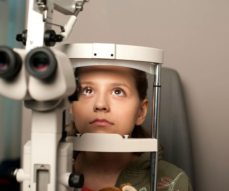
Specialist assessment & anatomical classification :
The initial assessment establishes where in the eye the inflammation is located, how active it is, and which structures are involved. A slit-lamp examination of the anterior segment is carried out alongside dilated fundoscopy and intraocular pressure measurement to look for the characteristic features of inflammation at each level — from anterior chamber cells and flare to vitreous haze, choroidal lesions, and macular involvement. The findings are used to classify the condition according to its anatomical subtype, which directly guides the investigation and treatment pathway that follows.
Investigation & identification of the underlying cause :
Once the pattern of inflammation has been established, a targeted set of investigations is arranged to identify or exclude an underlying cause. This typically includes relevant blood tests for autoimmune and infectious markers, chest imaging where sarcoidosis is a consideration, and advanced ocular imaging such as OCT and fluorescein angiography to assess the extent of posterior segment involvement. Where an infectious cause is suspected, aqueous sampling may be required. Where a systemic condition is identified or strongly suspected, liaison with the relevant medical specialty is arranged as part of a coordinated approach to the patient's overall care.
Medical treatment & inflammation control :
Treatment is selected based on the anatomical location of the inflammation, its underlying cause, and how severe the active episode is. Anterior uveitis is generally managed with topical corticosteroid drops and cycloplegic agents to suppress the inflammatory response and prevent complications such as posterior synechiae. More extensive or posterior inflammation typically requires periocular or intravitreal corticosteroid injections and, in many cases, systemic immunosuppressive therapy to achieve sustained disease control. Where standard immunosuppression proves insufficient, advanced biologic agents may be considered under close specialist supervision with regular monitoring of both ocular and systemic response.
Complication monitoring & long-term management :
Chronic or recurrent ocular inflammation carries a real risk of secondary complications — including cataract, raised intraocular pressure, cystoid macular oedema, and optic nerve damage — that can develop gradually and affect vision significantly if not detected early. Structured follow-up appointments include slit-lamp assessment, intraocular pressure monitoring, and OCT imaging of the macula and optic nerve to catch these changes at the earliest stage. Where surgical intervention becomes necessary, it is planned carefully with the inflammatory disease brought under optimal control beforehand to reduce the risk of post-operative recurrence.
Ocular inflammatory conditions are among the more complex presentations in ophthalmology, and managing them well requires both thorough investigation and consistent long-term attention. At Vinayaka Nethralaya, every patient with eye inflammation receives a careful initial assessment, an appropriate aetiological workup, and a treatment plan tailored to their specific condition. The goal throughout is to bring the inflammation under control as effectively as possible, prevent structural complications, and preserve the best achievable vision over the long term. Our team has been providing this level of specialist care to patients across Bengaluru since 1994.
What is included
Your complete eye inflammation care experience at our centre
Questions about eye inflammation treatment