
Glaucoma Treatment at Vinayaka Nethralaya
Protecting your vision from glaucoma through early detection & consistent long-term care
Glaucoma is one of the leading causes of irreversible vision loss worldwide, and what makes it particularly concerning is that it typically causes no pain and no noticeable symptoms in its early stages. By the time most patients become aware of a change in their vision, significant and permanent damage to the optic nerve has already taken place. The condition develops when the optic nerve — which carries visual information from the eye to the brain — is gradually damaged, most commonly as a result of raised pressure within the eye. Several distinct forms exist, including primary open-angle glaucoma, normal tension glaucoma, angle-closure glaucoma, and secondary glaucomas arising from other eye conditions. Each requires a different management approach. The single most effective way to prevent vision loss from glaucoma is to detect it early and bring intraocular pressure under consistent control. At Vinayaka Nethralaya, glaucoma assessment and management have been a central part of our clinical practice since 1994.
Optic nerve assessment and visual field monitoring
Clear, affordable consultation fees
Ophthalmologist with over four decades of glaucoma management experience
Glaucoma assessment & diagnosis
A thorough initial assessment is carried out to establish whether glaucoma is present, which subtype it represents, and how far it has progressed. This includes precise intraocular pressure measurement, gonioscopy to examine the drainage angle of the eye, detailed evaluation of the optic nerve head, and corneal thickness measurement to ensure pressure readings are interpreted accurately. A structured risk assessment is completed for each patient, taking into account age, family history, ethnicity, and disc morphology, to determine the appropriate level of monitoring and the urgency of treatment.
Optic nerve imaging & visual field assessment
Monitoring the optic nerve and visual field over time is central to understanding whether glaucoma is stable or progressing. OCT imaging of the retinal nerve fibre layer and optic nerve head provides objective, repeatable structural measurements that can detect early deterioration — often before any functional change is apparent to the patient. Visual field testing is performed at regular intervals to track the pattern and rate of any functional loss. Both sets of data are reviewed together to assess whether the current treatment is providing sufficient protection to the optic nerve.
Medical & laser treatment
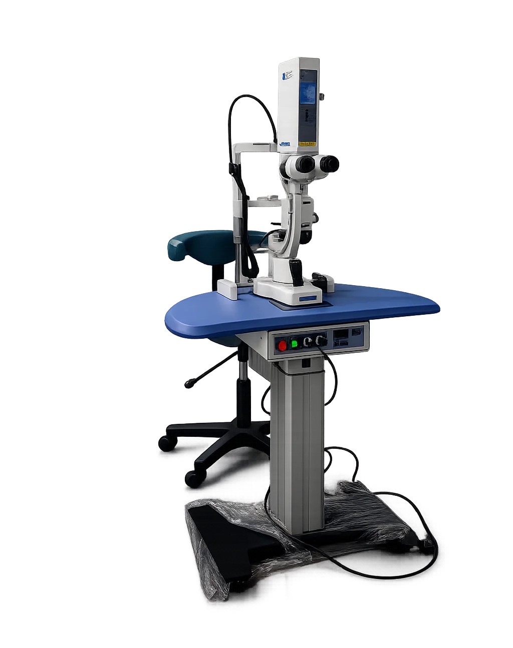
The primary aim of glaucoma treatment is to lower intraocular pressure to a level at which the optic nerve is protected from further damage. This is most commonly achieved initially through pressure-lowering eye drops, with additional agents introduced as needed to reach the target pressure. Selective laser trabeculoplasty is offered as an equally effective first-line option for open-angle glaucoma — it works by improving drainage through the trabecular meshwork and carries the advantage of removing the daily burden of drop instillation for patients who are suitable candidates.
Surgical management & long-term monitoring
When drops and laser treatment are not sufficient to halt progression, surgical intervention is considered. The choice of procedure depends on the glaucoma subtype and the degree of pressure reduction required. Minimally invasive options are appropriate for milder disease, while more advanced glaucoma may require trabeculectomy or tube shunt surgery to achieve a satisfactory target pressure. All patients who undergo surgery receive close post-operative follow-up with regular pressure checks, structural imaging, and visual field assessment to confirm that the surgical outcome is being maintained over time.
Glaucoma cannot be reversed — the damage already present at the time of diagnosis is permanent. What specialist-led care achieves is preventing further damage from occurring. At Vinayaka Nethralaya, glaucoma patients receive a thorough initial evaluation, an individually tailored treatment plan, and the kind of consistent long-term monitoring that this condition requires. Our aim is straightforward: to keep intraocular pressure under control, track the optic nerve carefully over time, and give every patient the best possible chance of retaining their functional vision for life.
What is included
Your complete glaucoma care experience at our centre
Questions about glaucoma treatment